The Cell Cycle
When living cells reach a certain size, they must divide or stop growing. Recall from an earlier module that cells are limited in size; this is because of their surface area to volume ratio. A number of cells in your body, such as nerve, red blood and muscle cells do not divide once they are mature. Other cells within multicellular organisms, such as skin or intestinal lining divide regularly to grow in size and/or repair tissues. In this module, we will focus on eukaryotic cell division for the purpose of repair and growth.
Watch the video clip of a fruit fly embryo dividing to increase in size by adding more cells.


Cell division is the organized process of creating new cells. Most cells do not divide constantly. Take a look at the diagram that represents the Cell Cycle, or a timeline of events that occur during the life of a cell in the image.
You can see in the diagram that the cell spends most of its life in interphase (green.) The cell may divide for growth or repair when signaled to do so. When the cell division occurs, it is composed of two highly organized events: mitosis and cytokinesis.
The cell is very busy during each of these stages. Learn about the events in each stage of the cell cycle.
![]() Stop and Think: Why would it be a bad to slice a cell in half haphazardly like a ninja? Why does a cell approach cell division in a neat, step-wise manner?
Stop and Think: Why would it be a bad to slice a cell in half haphazardly like a ninja? Why does a cell approach cell division in a neat, step-wise manner?
Organization of DNA
You learned in the notes above how important it was to properly distribute DNA to the two daughter cells resulting from cell division. One of the ways that the cell ensures the correct amount of DNA makes it into two cells is by being able to “package” DNA in different forms. Let’s learn about these different forms of DNA below:
Recall that DNA is a nucleic acid and one of the 4 types of macromolecules within cells. DNA is composed of monomer units called nucleotides.
When a eukaryotic cell is not reproducing, its DNA is inside the nucleus as disorganized, long strands called chromatin. Before a cell divides, the chromatin thickens and shortens into distinctly visible bodies called chromosomes. Each chromosome is a single DNA molecule. It is associated with proteins called histone proteins. Find each of these structures below:

In order for a cell to divide, its chromosomes must first make a copy of themselves (DNA replication) so that each new daughter cell gets the original amount of chromosomes.

Just after chromosome replication occurs, the two chromosomes (old one and new one) are stuck together. These 2 structures are referred to as sister chromatids. They are joined together by a central protein bundle called the centromere. The sister chromatids are exact copies of each other, and each one will be distributed to one of the new daughter cells.

Chromosome Number
Each species has a characteristic number of chromosomes in the nucleus of all of its cells. The number of chromosomes has nothing to do with the complexity of the organism. For instance, a chicken has 78 chromosomes, a black mulberry plant has 308, and the human chromosome number is 46.
Below is a picture, called a karyotype, of each of the 46 human replicated chromosomes. Humans have 2 sex chromosomes, X and Y, which determine the sex of the individual. XY is a male and XX is a female. Can you find these in the diagram below? The other 44 chromosomes are called autosomes, and they carry genes for all of the other traits of an individual.

The 46 human chromosomes are arranged in 23 homologous pairs so that there are two of each type of chromosome. Each chromosome of the pair is called a homolog, or homologous chromosome. Each homolog carries genes for a particular trait in the same place on its own chromosome. For this reason, every individual has two alternate forms of each gene that are referred to as alleles. Half of an individuals genetic information comes from the mother and the other half comes from the father.
Take a look at the Karyotype below. This particular karyotype shows a condition called trisomy 21, meaning that there are 3 of the 21st chromosome. An individual with trisomy 21 displays Down Syndrome.

Cell Regulation/Cancer
In a perfect world, cells can regulate their cycles so that the organism maintains a healthy rate of growth and repair. Cells do this in a number of ways by controlling what cells move through the cell cycle and when, by repairing damage to DNA and by stopping or slowing erratic growth.
Cancer is a disease where cells grow uncontrollably. Cancer cells can infiltrate and disrupt the growth of important tissues in the body with tumors, use blood supply and resources, and may ultimately cause death.
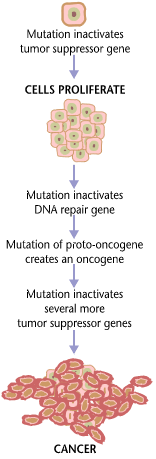
Cancer is caused by the mutations (or a change to DNA) of a combination of genes. Sometimes these mutations are inherited. Sometimes mutations are caused by mutagens in our environment. Take a look at the picture on the right that shows a possible progression of mutations that would cause cancer.
Sometimes cancer is described as hundreds of diseases rather than just one. This is because the cause of cancer can originate in different types of cells and at different genes within those cells that normally regulate the cell cycle correctly.
Watch the video below from NOVA’s “Battle in the War on Cancer: Breast Cancer.” It explains one of the ways in which cancer cells originate and move to other parts of the body.
While watching, think about the following questions:
- What is an oncogene and how is it dangerous for a cell?
- How does cancer spread? What characteristics of a cancer cell allow this?
![]()
(source)